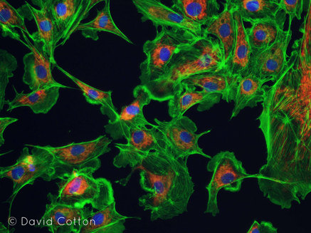
This image is one of my favourites from a residency I undertook earlier this year in New York (see previous post). Technically, it's three images, recombined to show cellular structures - the cytoskeleton has been dyed green, mitochondria in red and each nucleus lit up in blue.
Crafting images like these requires access to some pretty sophisticated equipment - an Olympus fluorescent microscope in this case, since you asked, a piece of kit I'm not so secretly coveting now - but far more than that I am indebted to the biotechnology staff (and students!) at JCC for their enthusiasm in sharing their research with me, their seemingly limitless knowledge of the field, and their trust in giving me almost free reign in the labs.
This image presents some interesting lines of inquiry. While these are fairly typical visualisations of cells, to my mind at least, they are also very selective in what they show - each filter reveals a different aspect of the cell, yet there is more invisible to us that isn't revealed by these three wavelengths of light. But in reality our vision is always constrained within a relatively narrow spectrum. There are an abundance of ultraviolet markings on flowers, for example, which insects are able to detect. What else are we missing?
 RSS Feed
